


Ultrasonography is based on imaging by sending sound waves to the body. The biggest difference from other imaging methods is that it does not contain radiation, that is, x-rays. It is used in the diagnosis of certain diseases, pregnancy follow-up and as an auxiliary imaging method for treatment.
With ultrasonography, some tissues and organs located in the inner region of the body that are not possible to be seen from the outside are displayed. This imaging method was obtained using high-frequency sound waves. It may be necessary to take ultrasonography by the physician during the diagnosis process of different diseases.
Ultrasonography is the examination of images that appear on the screen after high frequency sound waves are sent to the body. It is an imaging method that we use in the diagnosis and treatment process in medicine. It is a method that is frequently used in the field of radiology and offers great advantages to physicians in the diagnosis and follow-up of diseases.
Ultrasound image quality varies depending on the device used. In general, the image is reflected on the screen in black and white. This image is evaluated by the specialist physician. We can say that it is an important diagnostic and monitoring tool because it has a guiding nature in various situations.
Ultrasonography is used in various branches of medicine. We can list the main branches of these as follows:
Structures such as the hip bone can also be visualized by ultrasonography. For example, hip ultrasonography is performed for screening purposes for hip anomalies that may occur in infants. It is used safely in infants and children due to the fact that it does not emit radiation.
The ultrasound method is an extremely safe and effective imaging method. It should be noted that it is withdrawn at the request of physicians. If your doctor deems it necessary, he requests this examination and your ultrasound is taken in this direction. It does not require any preliminary preparation, except for some special cases. It is possible to be filmed and interpreted on the same day.
The importance of imaging methods in the medical field is great. They have a guiding and enlightening quality when diagnosing diseases. Since it provides imaging of organs and tissues that cannot be seen from the outside, the ultrasound method is also one of the extremely important imaging methods.
One of the most important advantages of ultrasound is that x-ray is not used. Unlike other imaging methods, it does not contain radiation. It takes advantage of sound waves. It does not have any harmful effects on pregnant women or babies. It is an effective and practical imaging method that is used safely.
The gestation period lasts about 40 weeks. During this period, the growth and development of babies should be monitored at regular December intervals. This condition is necessary for the health of the baby. In addition, parents are also happy to be able to see their children.
During pregnancy follow-up, ultrasound method is used by doctors who are Gynecologists and Obstetricians. When the last weeks of pregnancy are approaching, the follow-ups become more frequent. The absence of harm to the baby or the mother ensures that this method can be used safely during pregnancy follow-up.
Vaginal ultrasonography may be required for a better view of the gestational sac in the first weeks of pregnancy. In this method, the ultrasound probe is pushed inward through the vagina, resulting in a better image quality. However, as the weeks progress, sufficient images can also be provided with the ultrasound probe placed on the abdomen.
The ultrasound method is an imaging method that can be requested by the physician in the diagnosis of many diseases. It is often the first applied imaging method because it is safer than other methods, it can be taken without requiring preliminary preparation, and it is practical.
However, it should be remembered that this method alone may not be enough to diagnose each disease. Since your doctor has mastered what is needed in this regard, the examinations he requests are the most accurate ones. If an imaging method other than ultrasound is needed, your doctor will guide you in the most accurate way.
Ultrasonography method is often needed, especially for imaging of organs in the abdomen. It is possible to use the ultrasound method for imaging structures such as gallstones, gallbladder blockages, fatty liver, kidney diseases, cysts.
However, in such conditions, Magnetic Resonance (MRI) or Computed Tomography (CT) may be preferred if further examinations are required. Here, the decision rests entirely with the physician who manages the diagnosis and treatment process.
In general, it does not require any preparation process, but there may be some special cases. For example, for vaginal ultrasonography examination, it is requested that women are cramped to urinate. Such a condition is sought because the pelvic organs are better visualized when the bladder is full.
Or, your doctor may order an 8-hour fast to view some abdominal internal organs. If nothing is indicated to you when requesting an ultrasound scan, it means that you do not need to make preliminary preparations. You can go to your ultrasound appointment directly without any preparation.
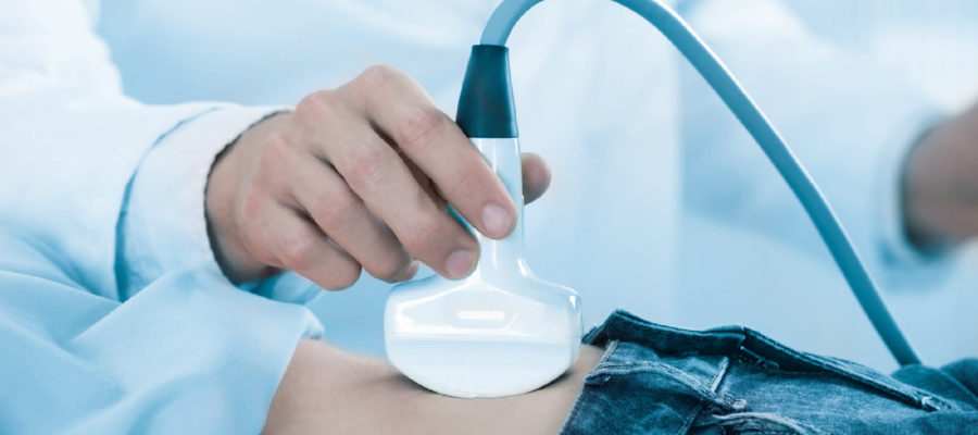
When applied over the abdomen, a gel is applied that will allow it to glide over the skin. Thus, the ultrasound probe is easily moved and the desired image is obtained by giving the right angle. The images obtained after the ultrasound scan can be printed out or just reported.
You can contact us immediately to get information about ultrasound prices 2026.