Fluoroscopy; is one of the oldest and most basic methods of radiology known. Its other name is röntgen, popularly known by the name Dec. It continues to be widely used today. It is used especially for the purpose of radiological examination of the digestive system, urinary tract, female reproductive organs and many parts of the body.
X-ray is used while patient examinations are performed. Structures that are not possible to be seen on normal films become visible by staining with drugs called contrast agents. In the content of drugs called contrast agents, there are barium and iodine radiopaque substances.
Contrast agents are given to the patient by drinking, enemas, with the help of a urinary catheter or intravenously, depending on the characteristics of the film to be examined. After the contrast agent is administered to the patient, the examined organ is monitored by the doctor on the screen and shots are taken in the positions deemed necessary.
The patient is involuntarily exposed to radiation during fluoroscopy. However, considering that the application is of vital importance for the patient, the side effects of a small amount of radiation are ignored Decently. As a result, the application is made taking into account the patient’s health.
The doctor or radiology technician performing fluoroscopy works behind a sheltered compartment or wears a lead apron to avoid constant radiation exposure. The patient is informed by the officer before the examination.
In addition, the patient takes off the clothes and metal materials on them before the shooting is done, and the patient wears a bib and the shooting is done. Patients with the possibility of pregnancy and the risk of allergies should inform the officer who will do the shooting about their condition before the shooting. After the end of the shooting process, the officer prepares a report and sends the report to the doctor who requested the examination.
1-Upper digestive system
2-Small intestines
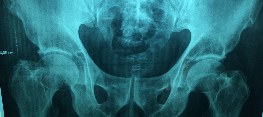
3-urinary tract
4-Female reproductive organs
It is called the esophagus-stomach-duodenum examination. For this examination, the patient is given a barium contrast agent to drink. With the introduction of barium contrast medium, the inner surface of the organs becomes visible. There are a number of preparatory stages before this examination is carried out.
At least 8 hours before the examination, the patient is not allowed to eat or even drink water and cigarettes. If the patient has medications that he is constantly using, it is allowed to drink with very little water under the supervision of a doctor who will perform fluoroscopy.
Immediately before the examination begins, the patient is given intravenous medication to restrict stomach movements. After that, the stomach swells and the patient is given a gas-making tablet to swallow in order to get a better image.
In addition, the patient is given a barium contrast drink and the progress of the drug through the esophagus and stomach is monitored by the doctor on the screen. Shots are performed in the amount and positions that the doctor considers necessary. Under normal conditions, an examination takes about half an hour. During this time, the patient does not feel any discomfort.
This examination is performed by making the patient drink a contrast agent. In some cases, it is performed by giving air to the patient through a tube that is swallowed.
During the 2 days before the examination, the patient is advised to take juicy foods. In addition, laxatives are given to the patient in terms of fasting and bowel cleansing during the 12-hour period before the examination.
During the examination, the patient is given a barium contrast drink and shots are taken in the required amount and in various positions until the drug enters the large intestine.
In cases where a detailed examination is required, a thin tube is swallowed by the patient through the nose and placed in the duodenum. Detailed shots can be made by giving air together with contrast agent from the tube. The duration of the examination varies according to the time of passage of the contrast agent into the large intestine.
In this examination, a contrast agent is introduced into the bladder with the help of a probe. Urinary tract imaging is performed with the patient’s urination. This procedure is called miction cystography. Before this procedure is performed, urine culture is performed in order to prevent a possible infection from spreading to the kidneys. The examination process is performed with the clean appearance of the urine Jul-ture.
In this examination, a probe is attached to the patient. The bladder is filled with the contrast medium given with the help of the probe and shots are made thanks to this. By removing the probe, the patient is allowed to urinate, and during this time, various shots are performed, examining whether there is a leak from the bladder. In this examination, the patient does not experience any discomfort except for inserting the probe and experiencing a feeling of tightness. This process takes approximately 25 minutes.
The uterus and tubes are examined by inserting a contrast agent into the uterus. This method is called hysterosalpingography. This method is performed 7-10 days after the beginning of the patient’s last menstruation. It is sufficient for the patient to take the pain relief tablet prescribed by the doctor before the examination.
The passage of the iodized contrast agent, which is given by inserting a thin tube into the uterus, from the uterus to the tubes and the abdominal cavity is monitored. Filming is carried out during this transition. This process takes about 10 minutes.
You can contact us immediately to get information about fluoroscopy prices 2024.